
Gum disease can develop without obvious symptoms. Early changes may remain difficult to notice. Dental imaging can reveal hidden changes. Bitewing images show upper and lower teeth together. They also show supporting bone levels. This makes Bitewing X-Rays useful.
Dentists can compare images during different visits. However, bitewing images cannot show everything. Severe bone loss may become visible. Other images may provide additional information. Your dentist combines imaging with examinations. This creates a more complete oral health picture.
How Bitewing X-Rays Can Show Bone Changes
Bone Levels Around Teeth
Supporting bone surrounds the roots of teeth. Healthy bone normally maintains a consistent height. Gum disease can gradually damage supporting tissues. Bone levels may then become lower. Bitewing images can reveal these changes. Dentists examine the bone near tooth surfaces.
They may compare images from different visits. This comparison can reveal changes over time. However, not every bone defect appears clearly. Image quality and positioning can affect visibility. Your dentist decides which images provide enough information.
Reveal Advanced Bone Loss
Severe gum disease can damage supporting bone. Those changes may appear on bitewing images. This makes imaging useful during dental evaluations. However, early disease may remain less obvious. Gum inflammation can begin before major bone loss.
Clinical examinations can detect other warning signs. Dentists may examine bleeding and gum depth. They also consider symptoms and medical history. A bitewing image adds another diagnostic view. It should not replace a complete gum examination.
Dentists Compare Changes
Dental imaging can become more useful over time. Previous images provide an important comparison point. Dentists can compare bone levels between visits. Changes may suggest progressing periodontal problems. Stable bone levels can also provide reassurance. However, comparisons require similar imaging techniques.
Your dentist considers differences before drawing conclusions. This makes professional interpretation especially important. Patients should avoid interpreting X-rays independently. A trained dentist can explain relevant findings clearly. This helps prevent unnecessary worry.
Show More Than Your Eyes
Some dental problems remain hidden during examinations. Teeth can look normal from outside. Bone changes can develop beneath the gums. Imaging helps dentists see these deeper structures. A dental bitewing X-ray can show useful details. It captures upper and lower teeth together.
It also provides information around supporting bone. However, different X-rays serve different purposes. Periapical images show roots more completely. Your dentist chooses imaging based on your needs.
Support Gum Diseas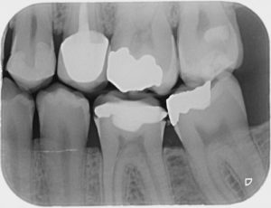 e Monitoring
Gum disease requires ongoing professional monitoring. Dentists assess changes during regular visits. Imaging can provide additional supporting information. Bitewing images may show changes around teeth. They can help track bone levels over time. However, gum measurements remain important too.
Dentists may check pockets around individual teeth. They can also assess bleeding and inflammation. Combining these findings provides better context. This approach supports more informed treatment decisions. Your dentist can explain what your images show.
Reveal Related Dental Problems
Bitewing images are not limited to bone. They commonly detect decay between teeth. They can also show certain dental abnormalities. Their focused view makes them useful routinely. Supporting bone can appear within these images. This provides additional information during examinations.
However, other problems need different imaging. Your dentist may recommend another X-ray type. The choice depends on your symptoms. It also depends on the suspected problem. Different images provide different diagnostic information.
Help Identify Changes Early
Early detection can simplify dental care. Finding changes sooner may support earlier treatment. However, bone loss often develops gradually. Regular dental examinations therefore remain important. Imaging can support these routine evaluations. Your dentist may recommend X-rays periodically.
Recommendations depend on your oral health needs. They also consider previous dental findings. Age and risk factors may influence recommendations. There is no single schedule for everyone. Your dentist can recommend appropriate imaging intervals.
Guide Further Dental Testing
A bitewing image may raise additional questions. Your dentist can then investigate those concerns. Additional X-rays may provide greater detail. Periapical images can show roots and surrounding bone. Periodontal measurements can also provide important information.
Together, these findings create a fuller picture. This prevents relying on one image alone. Your symptoms and dental history also matter. Your dentist considers all available evidence. This supports more appropriate diagnostic decisions.
Why Gum Disease Can Change What X-Rays Show
Gum Disease Can Affect Supporting Bone
Periodontal disease can damage supporting structures. These structures help keep teeth stable. Advanced inflammation can contribute to bone destruction. As bone decreases, teeth may lose support. Imaging can sometimes reveal these changes. The amount of visible loss can vary.
Early disease may not show clearly. Clinical examination remains necessary for proper diagnosis. Dentists consider both visible and hidden signs. This creates a more complete assessment. Prompt evaluation can help address concerning changes.
Bone Loss Can Happen Gradually
Gum disease usually does not damage bone overnight. Changes can develop gradually over time. This makes regular dental care important. Patients may notice few symptoms initially. Routine examinations can identify concerning changes sooner. X-rays can provide useful supporting information.
Dentists can compare images from previous visits. Gradual changes may become easier to recognize. However, imaging remains only one diagnostic tool. Your dentist considers your entire oral health picture. This includes symptoms and periodontal measurements.
What Should You Know Before Your X-Ray?
Your Dentist Decides When Imaging Helps
Not every patient needs identical X-rays. Dental imaging should support a clinical purpose. Your dentist considers your current oral health. Previous dental problems can influence recommendations. Symptoms may also require specific imaging.
Bitewing images are useful for certain concerns. Other situations may require different X-rays. Your dentist chooses the appropriate imaging type. This helps avoid unnecessary procedures. It also ensures important areas receive proper attention.
The Procedure Usually Takes Very Little Time
Bitewing imaging is generally quick and simple. A small sensor sits inside your mouth. You bite gently to hold it steady. The dentist then captures the image. Several images may be needed for coverage.
The process usually takes only a short time. Some patients may feel mild pressure. Tell your dental team if discomfort occurs. They can adjust the sensor position. Good positioning helps create clearer images.
Conclusion
Bitewing X-rays can reveal important bone changes. This becomes especially useful with severe gum disease. However, X-rays cannot diagnose every gum problem. Dentists combine imaging with clinical examinations.
They may also recommend other dental X-rays. Regular monitoring helps identify changes over time. If you have gum concerns, seek evaluation. Contact Nikahd Dental for professional dental care. Ask about Bitewing X-Rays in Corona during your visit.